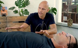
The Breatheology Method
Works on every dimension of the human body by uniting:
- Powerful breathing & lung training techniques
- Scientific knowledge on human physiology & neurology
- Proven visualization and meditation techniques
By combining these tools in the right way the Breatheology Method helps our students become conscious of their breathing to relax on demand and perform at the highest level.
Benefits
Optimize Performance – Increase Resilience – Better Sleep – Reduce Stress & Faster Recovery – Enhance Willpower – More Focus & Concentration – Transform Depression – Ease Anxiety – Faster Rehabilitation
… and much more breathing benefits


Free eBook & Breath Training
Start strengthening your respiratory muscles and build up your immune system.
- Free Online Breath Training Course
- The bestseller - "Breatheology - The art of Conscious Breathing" - Available in 11 languages
Languages: English, German, Portuguese, Chinese, French, Spanish, Arabic, Italian, Russian, Danish & Catalan
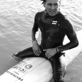
MUST READ: Stig Severinsen breaks new Guinness World Record
202.0 meters underwater in the open ocean on a single breath!


Start Your Journey Today
Begin or expand your breath work journey by choosing the course that is right for you.
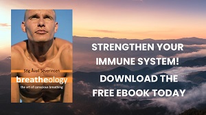
Free eBook & Breath Training
Strengthen your breath, body and mind with the free online Breath Training course and the free eBook
Breatheology ESSENTIALS™
Start learning the Breatheology Method at home through a series of easy instructional videos
Breatheology ADVANCED™
The Breatheology Course for improved relaxation, performance, and mental strength